

Description
| Reactivity: | Human, Mouse, Rat |
| Applications: | WB, FC, IC |
| Host Species: | Rabbit |
| Isotype: | IgG |
| Clonality: | Monoclonal antibody |
| Gene Name: | endonuclease G |
| Gene Symbol: | ENDOG |
| Synonyms: | Endonuclease G, mitochondrial; Endo G |
| Gene ID: | 2021 |
| UniProt ID: | Q14249 |
| Clone ID: | 25GB3035 |
| Immunogen: | A synthesized peptide derived from human Endo G |
| Dilution: | WB 1:1,000-1:5,000; FC 1:2,000; IC 1:100-1:1,000 |
| Purification Method: | Affinity purified |
| Concentration: | Lot dependent |
| Buffer: | PBS with 0.02% sodium azide, 50% glycerol, pH7.3. |
| Storage: | Store at -20°C. Avoid freeze/thaw cycles. |
Background
The protein ENDOG encoded by this gene is a nuclear encoded endonuclease that is localized in the mitochondrion. The encoded protein is widely distributed among animals and cleaves DNA at GC tracts. This protein is capable of generating the RNA primers required by DNA polymerase gamma to initiate replication of mitochondrial DNA.
Images
") |
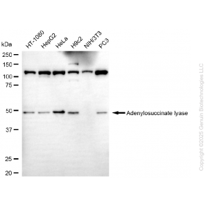
Western blotting analysis using anti-endonuclease G antibody (Cat#71216). Total cell lysates (30 μg) from various cell lines were loaded and separated by SDS-PAGE. The blot was incubated with anti-endonuclease G antibody (Cat#71216, 1:5,000) and HRP-conjugated goat anti-rabbit secondary antibody (Cat#201, 1:20,000) respectively. Image was developed using FeQ™ ECL Substrate Kit (Cat#226). |
") |
Western blotting analysis using anti-endonuclease G antibody (Cat#71216). Endonuclease G expression in wild type (WT) and endonuclease G (ENDOG) knockout (KO) HSHC cells with 20 μg of total cell lysates. Hsp90 α serves as a loading control. The blot was incubated with anti-endonuclease G antibody (Cat#71216, 1:5,000) and HRP-conjugated goat anti-rabbit secondary antibody (Cat#201, 1:20,000) respectively. Image was developed using FeQ™ ECL Substrate Kit (Cat#226). |
") |
Flow cytometric analysis of Endonuclease G expression in HepG2 cells using anti-Endonuclease G antibody (Cat#71216, 1:2,000). Green, isotype control; red, Endonuclease G. |
") |
Immunocytochemical staining of HepG2 cells with anti-Endonuclease G antibody (Cat#71216, 1:1,000). Nuclei were stained blue with DAPI; Endonuclease G was stained magenta with Alexa Fluor® 647. Images were taken using Leica stellaris 5. Protein abundance based on laser Intensity and smart gain: Medium. Scale bar, 20 μm. |


Reviews
There are no reviews yet.