

Description
| Reactivity: | Human, Mouse, Rat |
| Applications: | WB, FC, IC |
| Host Species: | Rabbit |
| Isotype: | IgG |
| Clonality: | Monoclonal antibody |
| Gene Name: | ribosomal protein SA |
| Gene Symbol: | RPSA |
| Synonyms: | SA; LBP; LRP; p40; uS2; 67LR; ICAS; lamR; 37LRP; LAMBR; LAMR1; LRP/LR; LBP/p40; NEM/1CHD4 |
| Gene ID: | 3921 |
| UniProt ID: | P08865 |
| Clone ID: | 25GB2950 |
| Immunogen: | A synthesized peptide derived from human 67kDa Laminin Receptor |
| Dilution: | WB 1:1,000-1:5,000; FC 1:2,000; IC 1:100-1:1,000 |
| Purification Method: | Affinity purified |
| Concentration: | Lot dependent |
| Buffer: | PBS with 0.02% sodium azide, 50% glycerol, pH7.3. |
| Storage: | Store at -20°C. Avoid freeze/thaw cycles. |
Background
Laminins, a family of extracellular matrix glycoproteins, are the major noncollagenous constituent of basement membranes. They have been implicated in a wide variety of biological processes including cell adhesion, differentiation, migration, signaling, neurite outgrowth and metastasis. Many of the effects of laminin are mediated through interactions with cell surface receptors. These receptors include members of the integrin family, as well as non-integrin laminin-binding proteins. This gene encodes a high-affinity, non-integrin family, laminin receptor 1. This receptor has been variously called 67 kD laminin receptor, 37 kD laminin receptor precursor (37LRP) and p40 ribosome-associated protein. The amino acid sequence of laminin receptor 1 is highly conserved through evolution, suggesting a key biological function. It has been observed that the level of the laminin receptor transcript is higher in colon carcinoma tissue and lung cancer cell line than their normal counterparts. Also, there is a correlation between the upregulation of this polypeptide in cancer cells and their invasive and metastatic phenotype. Multiple copies of this gene exist, however, most of them are pseudogenes thought to have arisen from retropositional events. Two alternatively spliced transcript variants encoding the same protein have been found for this gene.
Images
") |
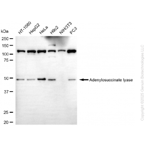
Western blotting analysis using anti-ribosomal protein SA antibody (Cat#71214). Total cell lysates (30 μg) from various cell lines were loaded and separated by SDS-PAGE. The blot was incubated with anti-ribosomal protein SA antibody (Cat#71214, 1:5,000) and HRP-conjugated goat anti-rabbit secondary antibody (Cat#201, 1:20,000) respectively. Image was developed using NaQ™ ECL Substrate Kit (Cat#716). |
") |
Western blotting analysis using anti-ribosomal protein SA antibody (Cat#71214). Ribosomal protein SA expression in wild type (WT) and ribosomal protein SA (RPSA) knockout (KO) HT-1080 cells with 20 μg of total cell lysates. Hsp90 α serves as a loading control. The blot was incubated with anti-ribosomal protein SA antibody (Cat#71214, 1:5,000) and HRP-conjugated goat anti-rabbit secondary antibody (Cat#201, 1:20,000) respectively. Image was developed using NaQ™ ECL Substrate Kit (Cat#716). |
") |
Flow cytometric analysis of Ribosomal protein SAc expression in HepG2 cells using anti-Ribosomal protein SA antibody (Cat#71214, 1:2,000). Green, isotype control; red, Ribosomal protein SA. |
") |
Immunocytochemical staining of HepG2 cells with anti-Ribosomal protein SA antibody (Cat#71214, 1:1,000). Nuclei were stained blue with DAPI; Ribosomal protein SA was stained magenta with Alexa Fluor® 647. Images were taken using Leica stellaris 5. Protein abundance based on laser Intensity and smart gain: Medium. Scale bar, 20 μm. |


Reviews
There are no reviews yet.