

Description
| Reactivity: | Human, Mouse, Rat |
| Applications: | WB, FC, IC |
| Host Species: | Rabbit |
| Isotype: | IgG |
| Clonality: | Monoclonal antibody |
| Gene Name: | transcription factor binding to IGHM enhancer 3 |
| Gene Symbol: | TFE3 |
| Synonyms: | TFEA; RCCP2; RCCX1; MRXSPF; bHLHe33 |
| Gene ID: | 7030 |
| UniProt ID: | P19532 |
| Clone ID: | 24GB2630 |
| Immunogen: | A synthesized peptide derived from human TFE3 |
| Dilution: | WB 1:1,000-1:5,000; FC 1:2,000; IC 1:100-1:1,000 |
| Purification Method: | Affinity purified |
| Concentration: | Lot dependent |
| Buffer: | PBS with 0.02% sodium azide, 50% glycerol, pH7.3. |
| Storage: | Store at -20°C. Avoid freeze/thaw cycles. |
Background
This gene encodes a basic helix-loop-helix domain-containing transcription factor that binds MUE3-type E-box sequences in the promoter of genes. The encoded protein promotes the expression of genes downstream of transforming growth factor beta (TGF-beta) signaling. This gene may be involved in chromosomal translocations in renal cell carcinomas and other cancers, resulting in the production of fusion proteins. Translocation partners include PRCC (papillary renal cell carcinoma), NONO (non-POU domain containing, octamer-binding), and ASPSCR1 (alveolar soft part sarcoma chromosome region, candidate 1), among other genes. Alternative splicing results in multiple transcript variants.
Images
") |
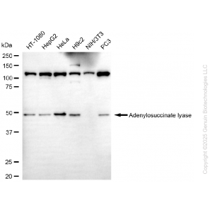
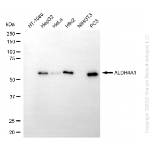
Western blotting analysis using anti-TFE3 antibody (Cat#69680). Total cell lysates (30 μg) from various cell lines were loaded and separated by SDS-PAGE. The blot was incubated with anti-TFE3 antibody (Cat#69680, 1:5,000) and HRP-conjugated goat anti-rabbit secondary antibody (Cat#201, 1:20,000) respectively. Image was developed using FeQ™ ECL Substrate Kit (Cat#226). |
") |
Western blotting analysis using anti-TFE3 antibody (Cat#69680). TFE3 expression in wild-type (WT) and TFE3 shRNA knockdown (KD) HeLa cells with 20 μg of total cell lysates. β-Tubulin serves as a loading control. The blot was incubated with anti-TFE3 antibody (Cat#69680, 1:5,000) and HRP-conjugated goat anti-rabbit secondary antibody (Cat#201, 1:20,000) respectively. Image was developed using anti-NaQ™ ECL Substrate Kit (Cat#716). |
") |
Flow cytometric analysis of TFE3 expression in H9c2 cells using anti-TFE3 antibody (Cat#69680, 1:2,000). Green, isotype control; red, TFE3. |
") |
Immunocytochemical staining of H9c2 cells with anti-TFE3 antibody (Cat#69680, 1:1,000). Nuclei were stained blue with DAPI; TFE3 was stained magenta with Alexa Fluor® 647. Images were taken using Leica stellaris 5. Protein abundance based on laser Intensity and smart gain: Medium. Scale bar: 20 μm. |


Reviews
There are no reviews yet.