

Description
| Reactivity: | Human |
| Applications: | WB |
| Host Species: | Rabbit |
| Isotype: | IgG |
| Clonality: | Monoclonal antibody |
| Gene Name: | H1.3 linker histone, cluster member |
| Gene Symbol: | H1-3 |
| Synonyms: | H1D; H1.3; H1F3; H1s-2; HIST1H1D |
| Gene ID: | 3007 |
| UniProt ID: | P16402 |
| Clone ID: | 25GB915 |
| Immunogen: | A synthesized peptide derived from human Histone H1.3 |
| Dilution: | WB 1:1,000-1:5,000 |
| Purification Method: | Affinity purified |
| Concentration: | Lot dependent |
| Buffer: | PBS with 0.02% sodium azide, 50% glycerol, pH7.3. |
| Storage: | Store at -20°C. Avoid freeze/thaw cycles. |
Background
Histones are basic nuclear proteins responsible for nucleosome structure of the chromosomal fiber in eukaryotes. Two molecules of each of the four core histones (H2A, H2B, H3, and H4) form an octamer, around which approximately 146 bp of DNA is wrapped in repeating units, called nucleosomes. The linker histone, H1, interacts with linker DNA between nucleosomes and functions in the compaction of chromatin into higher order structures. This gene is intronless and encodes a replication-dependent histone that is a member of the histone H1 family. Transcripts from this gene lack polyA tails but instead contain a palindromic termination element. This gene is found in the large histone gene cluster on chromosome 6.
Images
") |
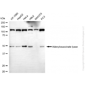
Western blotting analysis using anti-H1-3 antibody (Cat#65317). Total cell lysates (30 μg) from various cell lines were loaded and separated by SDS-PAGE. The blot was incubated with anti-H1-3 antibody (Cat#65317, 1:5,000) and HRP-conjugated goat anti-rabbit secondary antibody (Cat#201, 1:20,000) respectively. Image was developed using NaQ™ ECL Substrate Kit (Cat#716). |
") |
Western blotting analysis using anti-H1-3 antibody (Cat#65317). H1-3 expression in wild-type (WT) and H1-3 shRNA knockdown (KD) HeLa cells with 20 μg of total cell lysates. Hsp90 α serves as a loading control. The blot was incubated with anti-H1-3 antibody (Cat#65317, 1:5,000) and HRP-conjugated goat anti-rabbit secondary antibody (Cat#201, 1:20,000) respectively. Image was developed using FeQ™ ECL Substrate Kit (Cat#226). |


Reviews
There are no reviews yet.