

Description
| Reactivity: | Human, Mouse |
| Applications: | WB, FC, IC |
| Host Species: | Rabbit |
| Isotype: | IgG |
| Clonality: | Monoclonal antibody |
| Gene Name: | BCR activator of RhoGEF and GTPase |
| Gene Symbol: | BCR |
| Synonyms: | ALL; CML; PHL; BCR1; D22S11; D22S662 |
| Gene ID: | 613 |
| UniProt ID: | P11274 |
| Clone ID: | 24GB6945 |
| Immunogen: | A synthesized peptide derived from human Bcr |
| Dilution: | WB 1:1,000-1:5,000; FC 1:2,000; IC 1:100-1:1,000 |
| Purification Method: | Affinity purified |
| Concentration: | Lot dependent |
| Buffer: | PBS with 0.01% thimerosal, 50% glycerol, pH7.3. |
| Storage: | Store at -20°C. Avoid freeze/thaw cycles. |
Background
A reciprocal translocation between chromosomes 22 and 9 produces the Philadelphia chromosome, which is often found in patients with chronic myelogenous leukemia. The chromosome 22 breakpoint for this translocation is located within the BCR gene. The translocation produces a fusion protein which is encoded by sequence from both BCR and ABL, the gene at the chromosome 9 breakpoint. Although the BCR-ABL fusion protein has been extensively studied, the function of the normal BCR gene product is not clear. The unregulated tyrosine kinase activity of BCR-ABL1 contributes to the immortality of leukaemic cells. The BCR protein has serine/threonine kinase activity and is a GTPase-activating protein for p21rac and other kinases. Two transcript variants encoding different isoforms have been found for this gene.
Images
") |
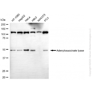
Western blotting analysis using anti-BCR antibody (Cat#63694). Total cell lysates (30 μg) from various cell lines were loaded and separated by SDS-PAGE. The blot was incubated with anti-BCR antibody (Cat#63694, 1:5,000) and HRP-conjugated goat anti-rabbit secondary antibody (Cat#201, 1:20,000) respectively. Image was developed using NaQ™ ECL Substrate Kit (Cat#716). |
") |
Western blotting analysis using anti-BCR antibody (Cat#63694). BCR expression in wild-type (WT) and BCR shRNA knockdown (KD) HeLa cells with 20 μg of total cell lysates. Hsp90 α serves as a loading control. The blot was incubated with anti-BCR antibody (Cat#63694, 1:5,000) and HRP-conjugated goat anti-rabbit secondary antibody (Cat#201, 1:20,000) respectively. Image was developed using NaQ™ ECL Substrate Kit (Cat#716). |
") |
Flow cytometric analysis of BCR expression in HepG2 cells using anti-BCR antibody (Cat#63694, 1:2,000). Green, isotype control; red, BCR. |
") |
Immunocytochemical staining of HepG2 cells with anti-BCR antibody (Cat#63694, 1:1,000). Nuclei were stained blue with DAPI; BCR was stained magenta with Alexa Fluor® 647. Images were taken using Leica stellaris 5. Protein abundance based on laser Intensity and smart gain: Medium. Scale bar, 20 μm. |


Reviews
There are no reviews yet.