

Description
| Reactivity: | Human, Mouse, Rat |
| Applications: | WB, FC, IC |
| Host Species: | Rabbit |
| Isotype: | IgG |
| Clonality: | Monoclonal antibody |
| Gene Name: | poly(A) binding protein nuclear 1 |
| Gene Symbol: | PABPN1 |
| Synonyms: | OPMD; PAB2; PABII; PABP2; PABP-2 |
| Gene ID: | 8106 |
| UniProt ID: | Q86U42 |
| Clone ID: | 24GB365 |
| Immunogen: | A synthesized peptide derived from human PABPN1 |
| Dilution: | WB 1:1,000-1:5,000; FC 1:2,000; IC 1:100-1:1,000 |
| Purification Method: | Affinity purified |
| Concentration: | Lot dependent |
| Buffer: | PBS with 0.02% sodium azide, 50% glycerol, pH7.3. |
| Storage: | Store at -20°C. Avoid freeze/thaw cycles. |
Background
This gene encodes an abundant nuclear protein that binds with high affinity to nascent poly(A) tails. The protein is required for progressive and efficient polymerization of poly(A) tails at the 3′ ends of eukaryotic transcripts and controls the size of the poly(A) tail to about 250 nt. At steady-state, this protein is localized in the nucleus whereas a different poly(A) binding protein is localized in the cytoplasm. This gene contains a GCG trinucleotide repeat at the 5′ end of the coding region, and expansion of this repeat from the normal 6 copies to 8-13 copies leads to autosomal dominant oculopharyngeal muscular dystrophy (OPMD) disease. Related pseudogenes have been identified on chromosomes 19 and X. Read-through transcription also exists between this gene and the neighboring upstream BCL2-like 2 (BCL2L2) gene.
Images
") |
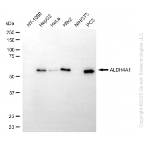
Western blotting analysis using anti-PABPN1 antibody (Cat#62337). Total cell lysates (30 μg) from various cell lines were loaded and separated by SDS-PAGE. The blot was incubated with anti-PABPN1 antibody (Cat#62337, 1:5,000) and HRP-conjugated goat anti-rabbit secondary antibody (Cat#201, 1:20,000) respectively. Image was developed using FeQ™ ECL Substrate Kit (Cat#226). |
") |
Western blotting analysis using anti-PABPN1 antibody (Cat#62337). PABPN1 expression in wild type (WT) and PABPN1 shRNA knockdown (KD) HT-1080 cells with 20 μg of total cell lysates. Hsp90 α serves as a loading control. The blot was incubated with anti-PABPN1 antibody (Cat#62337, 1:5,000) and HRP-conjugated goat anti-rabbit secondary antibody (Cat#201, 1:20,000) respectively. Image was developed using NaQ™ ECL Substrate Kit (Cat#716). |
") |
Flow cytometric analysis of PABPN1 expression in HepG2 cells using PABPN1 antibody (Cat#62337, 1:2,000). Green, isotype control; red, PABPN1. |
") |
Immunocytochemical staining of Hela cells with anti-PABPN1 antibody (Cat#62337, 1:1,000). Nuclei were stained blue with DAPI; PABPN1 was stained magenta with Alexa Fluor® 647. Images were taken using Leica stellaris 5. Protein abundance based on laser Intensity and smart gain: High. Scale bar: 20 μm. |


Reviews
There are no reviews yet.